
Pulmonic Valve Disease in Dogs: Symptoms, Types, Causes, Signs & Prevention

Canine Bible is reader-supported. We receive affiliate commissions via some of our links. Learn more.
A comprehensive guide to pulmonic valve disease in dogs, including causes, predisposed breeds, warning signs, diagnostic testing, disease severity, treatment options, prognosis and what pet parents should know when caring for a dog with this congenital heart condition.
Pulmonic Valve Disease at a Glance
Most common in
Young dogs and predisposed breeds such as Bulldogs, Boxers and Beagles.
Common signs
Heart murmur, fatigue, exercise intolerance, weakness and fainting.
Diagnosis
Cardiac exam, echocardiogram, Doppler ultrasound and ECG.
Treatment
Monitoring, medications and balloon valvuloplasty when appropriate.
Prognosis
Often good in mild cases; severe disease carries greater cardiac risk.
Congenital cases
Pulmonic stenosis represents about 20%–35% of congenital heart defects in several studies.
What Is Pulmonic Valve Disease in Dogs?
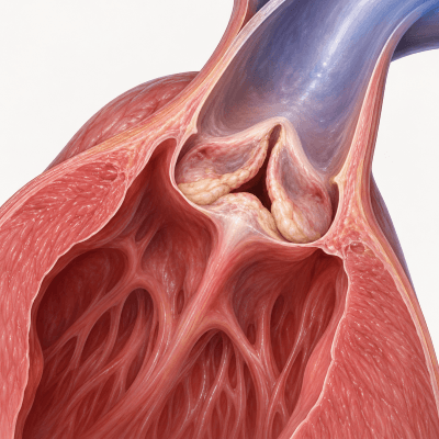
Pulmonic valve disease in dogs most commonly refers to pulmonic stenosis, a congenital heart defect in which the pulmonic valve or nearby outflow tract is abnormally narrowed. The pulmonic valve normally allows blood to move from the right ventricle into the pulmonary artery and toward the lungs. When the opening is narrowed, the right side of the heart must generate greater pressure to push blood through it. Over time, this increased workload can cause thickening and enlargement of the right ventricle and may contribute to exercise intolerance, weakness, fainting, abnormal heart rhythms or, in severe cases, right-sided congestive heart failure. Mildly affected dogs may show no outward symptoms and may only be identified after a veterinarian detects a heart murmur during a routine examination.


Types
Pulmonic Valve Disease in dogs can take several forms depending on where the narrowing or abnormality occurs. Some affect the pulmonic valve itself, while others involve the area just below or above the valve. Understanding the different types can help pet parents recognize why severity, treatment options, and prognosis may vary from one dog to another.
Valvular Pulmonic Stenosis
The most common form, caused by abnormal or narrowed pulmonic valve leaflets that restrict blood flow from the heart.
Type A Pulmonic Stenosis
Characterized by fused or thickened valve leaflets with a relatively normal-sized pulmonic valve opening.
Type B Pulmonic Stenosis
Involves thicker, less mobile valve tissue and an abnormally small valve opening, often creating more complex obstruction.
Subvalvular Pulmonic Stenosis
A less common form in which narrowing develops below the pulmonic valve within the right ventricular outflow tract.
Supravalvular Pulmonic Stenosis
A rare form in which the obstruction occurs above the pulmonic valve within the pulmonary artery.


Pulmonic Valve Dysplasia
A congenital malformation in which the valve leaflets are abnormally thickened, misshapen or poorly mobile.
Pulmonic Regurgitation
Occurs when the pulmonic valve does not close completely, allowing some blood to leak backward into the right ventricle.
Combined Congenital Heart Defects
Pulmonic valve abnormalities may occur alongside other congenital heart defects, which can increase cardiac strain and complicate treatment.
Predisposed Dog Breeds
While Pulmonic Valve Disease can affect any dog, certain breeds have a higher genetic risk. Inherited heart abnormalities and breed-related predisposition make some dogs more likely to develop pulmonic stenosis and other pulmonic valve defects than others.
| Size / Group | Breeds | Notes on Risk Factors |
|---|---|---|
| Brachycephalic Breeds | English Bulldog, French Bulldog, Boxer | Among the breeds most frequently associated with pulmonic stenosis. Congenital valve abnormalities and inherited predisposition may substantially increase risk. |
| Hound & Sporting Breeds | Beagle, Boykin Spaniel, Cocker Spaniel | Certain lines show an increased occurrence of congenital pulmonic stenosis, suggesting an important hereditary component. |
| Terrier Breeds | West Highland White Terrier, Scottish Terrier, Staffordshire Bull Terrier | Several terrier breeds have been reported more frequently among dogs diagnosed with congenital pulmonic valve narrowing. |
| Small & Toy Breeds | Chihuahua, Miniature Schnauzer | Congenital valve malformations can occur in smaller breeds, although overall risk varies between breeds and individual bloodlines. |
| Working & Northern Breeds | Mastiff, Bullmastiff, Samoyed | Breed-associated genetic factors may increase susceptibility to abnormal development of the pulmonic valve or right ventricular outflow tract. |
| Other Dogs | Mixed-Breed Dogs and Other Purebred Dogs | Pulmonic valve disease can occur in any dog, even without a recognized breed predisposition. A persistent heart murmur in a puppy or young dog should be evaluated by a veterinarian. |
Causes
Pulmonic Valve Disease in dogs usually develops because of a congenital abnormality in the pulmonic valve or nearby heart structures, often with an inherited component in predisposed breeds. Rather than developing from lifestyle factors, the condition is typically present from birth. Understanding the genetic and developmental factors involved can help pet parents make informed decisions about screening, breeding, early diagnosis, and long-term cardiac care.
Congenital Development
Pulmonic valve disease is usually present from birth because the valve or nearby outflow tract does not develop normally.
Genetics
Inherited factors are believed to increase risk in predisposed breeds such as Bulldogs, Boxers, Beagles, and some terriers.
Pulmonic Valve Dysplasia
Abnormally thickened, misshapen, or poorly mobile valve leaflets can restrict blood flow from the right ventricle.
Valve Leaflet Fusion
Partial fusion of the pulmonic valve leaflets can narrow the opening and create an obstruction to normal blood flow.
Annular Hypoplasia
Some dogs are born with an abnormally small pulmonic valve opening, limiting the amount of blood that can pass through.
Subvalvular or Supravalvular Narrowing
Less commonly, obstruction develops below the pulmonic valve or above it within the pulmonary artery rather than at the valve itself.
Associated Congenital Abnormalities
Pulmonic stenosis can occur alongside other structural heart abnormalities, including unusual coronary artery anatomy in some dogs.
Symptoms
Pulmonic Valve Disease symptoms in dogs can vary widely depending on the severity of the narrowing, blood flow obstruction, and strain on the right side of the heart. Some dogs may show no obvious signs at first, while others develop more noticeable symptoms as the condition becomes more severe. Common warning signs include a heart murmur, exercise intolerance, fatigue, weakness, fainting, rapid or labored breathing, and poor growth in puppies. Because many of these symptoms can also be caused by other heart or respiratory conditions, any persistent or unusual changes should be evaluated by a veterinarian.
- Heart murmur
- Exercise intolerance
- Reduced stamina or easy fatigue
- Weakness
- Fainting or collapse
- Rapid or labored breathing
- Poor growth in puppies
- Abnormal heart rhythms
- Abdominal swelling in severe cases
- Signs of right-sided heart failure
Pulmonic valve disease primarily affects the heart but can also influence circulation, breathing, stamina, and overall exercise tolerance.
Stages & Progression of Pulmonic Valve Disease in Dogs
Pulmonic Valve Disease in dogs can vary in severity depending on the degree of valve narrowing, the pressure overload on the right side of the heart, and whether complications such as arrhythmias or heart failure develop. Staging helps veterinarians determine how severe the disease is, how much strain it is placing on the heart, and which treatment options may be most appropriate. Understanding the stage of disease can also provide important insight into prognosis, expected progression, and the level of monitoring or intervention your dog may need.
Mild Disease
Dogs may show no outward symptoms. A heart murmur may be the only finding, and Doppler pressure gradients are typically below 50 mmHg.
Moderate Disease
Pressure across the pulmonic valve increases, often around 50–80 mmHg. Some dogs may develop fatigue, exercise intolerance, weakness, or occasional fainting.
Severe Disease
Pressure gradients above about 80 mmHg indicate severe obstruction. The right ventricle works much harder and heart enlargement, fainting, or arrhythmias may occur.
Advanced Complications
Severely affected dogs may develop significant right-sided heart enlargement, abnormal rhythms, fluid buildup, reduced exercise capacity, or right-sided congestive heart failure.
Pulmonic valve disease does not always progress through predictable stages over time. Mild, moderate, and severe categories are generally based on Doppler measurements and overall cardiac findings, while advanced complications describe the effects that may develop in severely affected dogs.
Top Ways to Prevent Pulmonic Valve Disease in Dogs
Because pulmonic valve disease is usually a congenital and potentially inherited heart defect, it cannot always be prevented. However, responsible breeding, cardiac screening of predisposed dogs, and regular veterinary checkups can help reduce hereditary risk and support early detection. Identifying heart murmurs and structural abnormalities early gives affected dogs the best chance for timely monitoring, treatment, and a better quality of life.
| Prevention Strategy | Why It Helps | How to Do It |
|---|---|---|
| Responsible Breeding | Pulmonic valve disease is usually congenital and may have an inherited component in predisposed breeds. | Avoid breeding dogs diagnosed with pulmonic stenosis or other significant congenital heart defects. |
| Screen Breeding Dogs | Cardiac screening may identify heart murmurs or structural abnormalities before affected dogs are bred. | Have breeding dogs examined by a veterinarian and consider echocardiography or cardiology screening in high-risk breeds. |
| Early Puppy Heart Exams | Pulmonic stenosis is present from birth, and a heart murmur may be detected before symptoms become obvious. | Schedule routine puppy examinations and ask your veterinarian to investigate persistent or significant heart murmurs. |
| Prompt Echocardiography | Early diagnosis helps determine the severity of valve narrowing and whether treatment or closer monitoring is needed. | Seek a veterinary cardiology evaluation if a murmur, fainting, weakness, or exercise intolerance is detected. |
| Follow Exercise Recommendations | Excessive exertion may increase cardiac demand in dogs with moderate or severe pulmonic stenosis. | Follow your veterinarian’s activity guidelines and avoid intense exercise if restrictions are recommended. |
| Maintain a Healthy Weight | Excess body weight can increase overall cardiovascular workload and make exercise more difficult. | Feed measured portions, limit excess treats, and maintain an appropriate body condition under veterinary guidance. |
| Regular Cardiology Monitoring | Follow-up testing can detect changes in pressure gradients, right-sided heart enlargement, or rhythm abnormalities. | Schedule repeat examinations, echocardiograms, or ECGs at the intervals recommended by your veterinary cardiologist. |
| Treat Significant Disease Early | Timely intervention may reduce pressure on the right side of the heart and lower the risk of serious complications. | Discuss medications or balloon valvuloplasty with a veterinary cardiologist when moderate or severe disease is diagnosed. |
Actionable Resources
If you want to support your dog’s heart health and manage Pulmonic Valve Disease, here are some resources to explore under veterinary guidance.
How Is Pulmonic Valve Disease In Dogs Diagnosed
Diagnosing pulmonic valve disease in dogs requires a comprehensive veterinary cardiac evaluation to confirm the abnormality, measure the severity of the obstruction, and assess its effects on the heart. Key steps include:
1. Physical Examination
Your veterinarian listens for a heart murmur and evaluates heart rate, pulse quality, breathing, gum color, exercise tolerance, and other signs of cardiovascular disease.
2. Echocardiogram
Cardiac ultrasound allows a veterinarian or cardiologist to examine the pulmonic valve, right ventricle, pulmonary artery, and surrounding structures for congenital abnormalities.
3. Doppler Ultrasound
Doppler imaging measures blood-flow velocity through the pulmonic valve and estimates the pressure gradient across the obstruction, helping classify disease as mild, moderate, or severe.
4. Electrocardiogram (ECG)
An ECG records the heart’s electrical activity and can identify abnormal rhythms or electrical patterns associated with right-sided heart enlargement and increased cardiac strain.
5. Chest X-Rays
Thoracic radiographs may show enlargement of the right side of the heart, changes in the pulmonary artery, or signs of fluid accumulation if heart failure has developed.
6. Advanced Cardiac Testing
CT imaging, angiography, cardiac catheterization, or other specialized testing may be recommended in complex cases to evaluate coronary anatomy, confirm unusual obstruction, or plan procedures such as balloon valvuloplasty.
Early detection is important because Pulmonic Valve Disease can place increasing strain on the right side of the heart—identifying the condition early allows for appropriate monitoring, timely treatment, and better management of potential complications and quality of life.
Nutritional Support & Supplements
Proper nutrition can play an important supportive role for dogs with Pulmonic Valve Disease by helping maintain healthy body weight, muscle condition, cardiovascular function, and overall quality of life. Depending on your dog’s needs and disease severity, veterinarians may recommend strategies such as balanced heart-supportive diets and supplements including omega-3 fatty acids, taurine, L-carnitine, CoQ10, or carefully managed sodium intake when appropriate.
Important: Nutrition and supplements cannot correct a narrowed pulmonic valve or replace cardiac treatment.
Taurine, L-carnitine, potassium, magnesium, sodium restriction, and other supplements may not be appropriate for every dog.
Always discuss dietary changes and supplements with your veterinarian or veterinary cardiologist, especially if your dog takes heart medications.
Supportive care—such as pain management, nutritional support, and lifestyle adjustments—plays a crucial role in maintaining comfort and quality of life during treatment. These options are not a cure for cancer and may interact with chemotherapy or other medications, so any diet or supplement plan should be reviewed with your veterinarian or veterinary oncologist.
Over-the-Counter Products
Over-the-counter treatments and supplements mentioned in this article for cancer in dogs can be found on Amazon and Chewy.



Always consult your veterinarian before starting any new treatment, medication, or home-care regimen for canine pulmonic valve disease. Puppies and dogs with persistent vomiting, diarrhea, weakness, or dehydration may require prompt veterinary care. If you can’t reach your vet, you can chat live with a registered online veterinary professional via our online vet chat or video chat support (24 hours a day, 7 days a week). Or use Chewy’s online vet services (6 a.m. – midnight ET).
Treatment & Care
According to current veterinary cardiology guidance, the most effective treatment and care for Pulmonic Valve Disease in dogs depends on the severity of the valve narrowing and its effect on the heart. Management may include ongoing monitoring for mild cases, medications such as beta-blockers in selected dogs, and balloon valvuloplasty for moderate to severe pulmonic stenosis to improve blood flow and reduce pressure on the right side of the heart. In more advanced cases, treatment may also involve managing arrhythmias, exercise intolerance, or signs of right-sided heart failure to help maintain comfort and quality of life.
Medications
Common medications used to treat pulmonic valve disease in dogs include:
| Medication / Class | Examples | How It Is Used | Important Considerations |
|---|---|---|---|
| Beta-Blockers | Atenolol | May be prescribed in selected dogs to slow the heart rate, reduce cardiac workload, and help control certain abnormal rhythms. | Not required for every dog with pulmonic stenosis. Heart rate, blood pressure, and clinical response should be monitored by a veterinarian or veterinary cardiologist. |
| Antiarrhythmic Medications | Sotalol, mexiletine, other veterinarian-selected drugs | Used when clinically significant ventricular or other abnormal heart rhythms develop. | Drug selection depends on the type of arrhythmia. ECG or Holter monitoring may be recommended to evaluate treatment response. |
| Diuretics | Furosemide | May be used if severe pulmonic valve disease progresses to congestive heart failure with abnormal fluid accumulation. | Can affect hydration, kidney function, and electrolyte levels. Bloodwork and clinical monitoring are important during treatment. |
| Aldosterone Antagonists | Spironolactone | May be added to heart-failure treatment in selected dogs to help manage fluid retention and the effects of chronic cardiac disease. | Kidney function and electrolyte levels, particularly potassium, may require monitoring. |
| Cardiac Support Medications | Pimobendan | May be considered in selected dogs that develop impaired cardiac function or congestive heart failure. | It does not correct pulmonic valve narrowing and is not routinely required for uncomplicated pulmonic stenosis. Use depends on the individual dog’s cardiac findings. |
| ACE Inhibitors | Enalapril, benazepril | May occasionally be incorporated into treatment plans when congestive heart failure or other cardiovascular complications are present. | Blood pressure, kidney values, and electrolytes should be monitored. These medications do not relieve the valve obstruction itself. |
| Emergency Supportive Therapy | Oxygen therapy and veterinarian-selected emergency medications | May be required during episodes of severe breathing difficulty, collapse, arrhythmia, or decompensated heart failure. | Emergency signs require immediate veterinary care and should not be managed with medications at home without professional guidance. |
| Balloon Valvuloplasty | Catheter-based balloon dilation | Although not a medication, it is the principal interventional treatment for many suitable dogs with moderate-to-severe valvular pulmonic stenosis. | The procedure directly reduces the obstruction, unlike medications. Suitability depends on valve anatomy, disease severity, and veterinary cardiology assessment. |
The best way to get these medications is at Chewy Pet Pharmacy:
Alternative & Integrative Therapies
Integrative and supportive therapies can help support overall comfort, reduce stress, and improve quality of life for dogs with Pulmonic Valve Disease. Options such as controlled low-impact activity, weight management, acupuncture, nutritional support, and stress reduction may be used alongside conventional cardiac care, but they do not correct the narrowed pulmonic valve and should always be discussed with your veterinarian or veterinary cardiologist to ensure they are safe and appropriate for your dog.
Surgical Options
Interventional or surgical treatment is one of the most important options for dogs with moderate to severe Pulmonic Valve Disease, especially when the valve narrowing significantly restricts blood flow and increases pressure on the right side of the heart. Depending on the valve anatomy and disease severity, treatment may involve balloon valvuloplasty to widen the narrowed valve opening, improve circulation, and reduce cardiac strain. In selected or more complex cases, specialized cardiac procedures may be considered if balloon intervention is not appropriate or if other congenital abnormalities are present. Your veterinarian or veterinary cardiologist will determine whether intervention is appropriate based on your dog’s anatomy, pressure gradient, symptoms, and overall health.
Emotional & Lifestyle Impact
A diagnosis of Pulmonic Valve Disease can be emotionally challenging for both dogs and their families. Dogs may experience reduced stamina, exercise intolerance, weakness, fainting, or activity limitations, while more severe cases may require medications, cardiac procedures, or ongoing specialist care. With thoughtful management, regular monitoring, and appropriate treatment, many affected dogs can continue to enjoy happy, comfortable lives while maintaining safe levels of activity and quality of life.
For Dogs
- May tire more quickly during walks or play
- Moderate or severe cases may require exercise restrictions
- Weakness or fainting can make strenuous activity unsafe
- Regular cardiology visits may become part of their routine
- Many dogs can still enjoy gentle play, affection, and family time
For Pet Parents
- Cardiac diagnoses can bring uncertainty about long-term health
- Specialist visits and testing can require added time and expense
- Activity may need to be adjusted based on disease severity
- Monitoring fainting, breathing, and stamina becomes important
- Following a clear cardiology plan can make daily care easier
A heart condition may change how your dog exercises, but it does not take away the joy of gentle play, companionship, and everyday moments together.
Better days.
Dog Pulmonic Valve Disease Prognosis
A dog’s Pulmonic Valve Disease prognosis depends on several factors, including the severity of valve narrowing, pressure gradient, presence of symptoms, degree of right-sided heart enlargement, abnormal heart rhythms, and response to treatment. Dogs with mild disease may live normal or near-normal lives, while severe cases have a greater risk of fainting, heart failure, or other cardiac complications. Working closely with your veterinary cardiology team can help guide treatment decisions, monitor progression, and maintain the best possible quality of life for your dog.
Key Factors That Influence Prognosis
Typical Annual Cost of Treating Canine Pulmonic Valve Disease
| Category | Estimated Cost Range | Notes |
|---|---|---|
| Initial Cardiology Evaluation | $250 – $600+ | Includes a veterinary cardiology consultation, physical examination, heart auscultation, and review of symptoms and medical history. |
| Echocardiogram & Doppler Testing | $400 – $1,200+ | Used to confirm pulmonic stenosis, examine valve anatomy, and measure blood-flow velocity and pressure gradients. |
| ECG, X-Rays & Additional Testing | $200 – $1,000+ | May include electrocardiography, chest X-rays, bloodwork, Holter monitoring, or advanced cardiac imaging when needed. |
| Cardiac Medications | $100 – $1,000+ per year | May include beta-blockers, antiarrhythmics, diuretics, or other cardiac medications depending on symptoms and complications. |
| Balloon Valvuloplasty | $3,000 – $8,000+ | A specialized catheter-based procedure used in suitable dogs with moderate-to-severe pulmonic stenosis. Usually a one-time procedural cost rather than an annual expense. |
| Advanced Cardiac Procedures | $5,000 – $12,000+ | Complex cases may require specialized balloon techniques, hybrid procedures, or surgery at an advanced veterinary cardiology center. |
| Follow-Up Cardiology & Imaging | $400 – $1,500+ per year | Repeat echocardiograms, ECGs, examinations, and bloodwork may be recommended to monitor cardiac changes and treatment response. |
| Emergency / Heart Failure Care | $1,000 – $5,000+ | Hospitalization, oxygen therapy, imaging, medications, and intensive monitoring may be needed if severe complications develop. |
| Nutritional Support & Supplements | $200 – $1,000+ per year | May include heart-supportive diets, omega-3 fatty acids, or other veterinarian-recommended nutritional support. |
| Pet Insurance Coverage | Varies | Coverage depends heavily on the policy. Congenital or previously diagnosed heart disease may be excluded as a pre-existing condition. |
| Typical Cost Range | Monitoring / Medical Management: $1,000 – $4,000+ per year | With Balloon Valvuloplasty: $4,000 – $10,000+ | |
Pulmonic Valve Disease care can add up quickly — from daily medications to possible surgeries. Pet insurance can ease the financial burden and ensure your dog gets the best treatment without compromise. If you want to compare plans and find out which providers offer the best coverage for pulmonic valve disease and chronic conditions, check out our full guides to the Best Pet Insurance for Dogs and Best Pet Wellness Plans.
Related Health Conditions
Some health conditions are closely linked to cancer because they may represent specific tumor types, affect similar organs, or cause overlapping symptoms. Understanding these related conditions can help dog owners recognize potential warning signs, learn how different cancers behave, and better understand the diagnostic and treatment options their veterinarian may recommend.
- Congenital Heart Defects
- Aortic Stenosis
- Tricuspid Valve Disease
- Arrhythmia
- Heart Failure
- Dilated Cardiomyopathy (DCM)
- Pulmonary Hypertension
- Right-Sided Congestive Heart Failure
Frequently Asked Questions
The Bottom Line
Pulmonic Valve Disease in dogs is a significant congenital heart condition, but with early detection, regular cardiology monitoring, and appropriate treatment, many affected dogs can continue to enjoy a good quality of life. Mild cases may require little more than routine follow-up, while moderate to severe disease may benefit from medications, exercise adjustments, or procedures such as balloon valvuloplasty. By recognizing warning signs such as exercise intolerance, fainting, weakness, or breathing changes and working closely with your veterinary team, you can help protect your dog’s heart health and comfort. Above all, consistent care, timely intervention, and thoughtful lifestyle management can make a meaningful difference throughout your dog’s life.